In Georgia, a fully automated pathomorphological laboratory functions in our Institute. It provides cytological, common pathomorphological, and immunohistochemical examination.
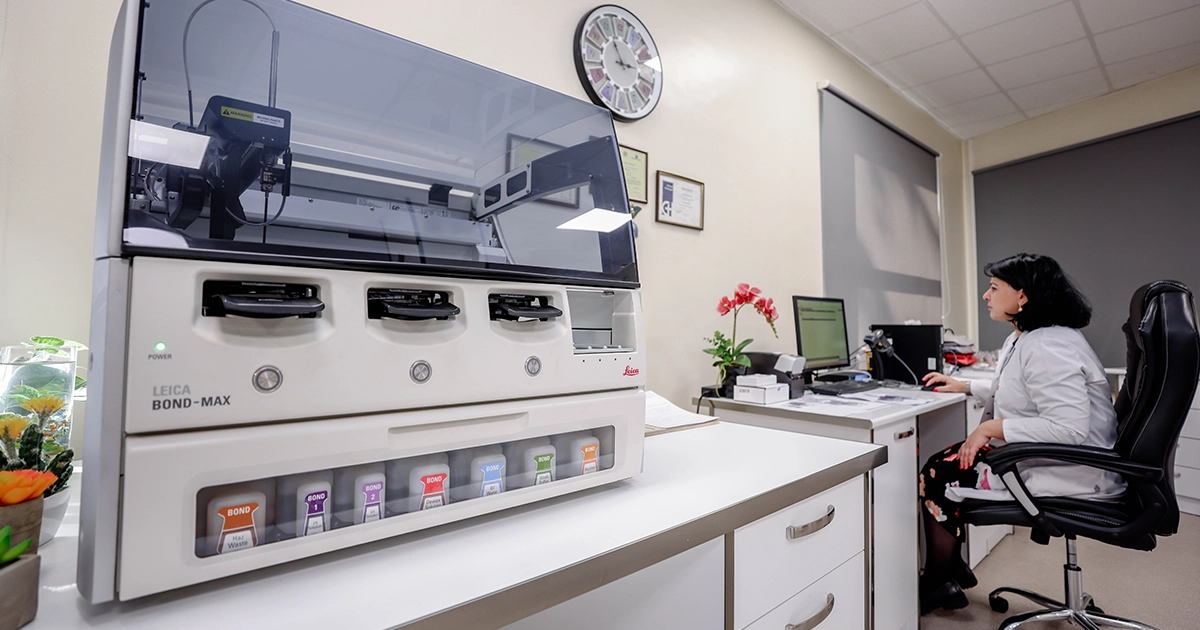
The Unit is equipped with modern apparatus and digital microscopes of the company LEICA. Fully automated immunohistochemical system - the latest model of LEICA AUTOSTAINER BOND MAX -2017 5th line. Microtom - LEICA RM 2255- MICROTOME. Tissue Processor – LEICA HistoCore PEARL latest model of 2017, and other subsidiary equipment of company LEICA, necessary for the diagnostics.
Benefits of pathomorphological laboratory
Key areas of the pathomorphological laboratory operation:
• Lifetime morphological diagnostics of pathological processes of various organs and systems.
• Clinical-anatomic analysis, prognostic assessment of processes
• Immunohistochemical examination is performed at AUTOSTAINER BOND MAX of LEICA company using a wide range of antibodies.
This method is used for: • Specifying tumor histogenesis. • Defining the primary locus according to the metastatic lesion • Determination of tumor anaplasia quality
• Determination of immunophenotype in lymphatic proliferation diseases
• Determination of estrogen and progesterone receptors and oncogene C-erbB-2/Her- 2/new in breast cancer cases. • Determination of tumor tissue sensitivity to hormonal and chemotherapeutic drugs.
Dont miss Koi news


