Ultrasonography (ultrasound examination) – is a method for examining internal organs and soft tissues by using ultrasound waves.
The procedure is safe not only for adults, but also for kids, and is also applied during pregnancy, as it is harmful for the fetus and does not have a radiation effect on the human body.
Purpose of Ultrasonography
Ultrasonography is frequently used in the very early stages for diagnostics of diseases and disorders in various organs or soft tissues as it has no side effects, has no harmful effect on the body, and is performed quickly - usually in less than half an hour.
Benefits of Ultrasonography:
It should be noted that ultrasound examination does not always give a clear image of the tissue in obese people.
Ultrasound can't be transmitted and is reflected from surfaces like bone and lesions with similar density, as well as gases that make it impossible to distinguish the structures beneath them.
Ultrasound is used to examine:
In addition, ultrasonography is used for the following purposes:
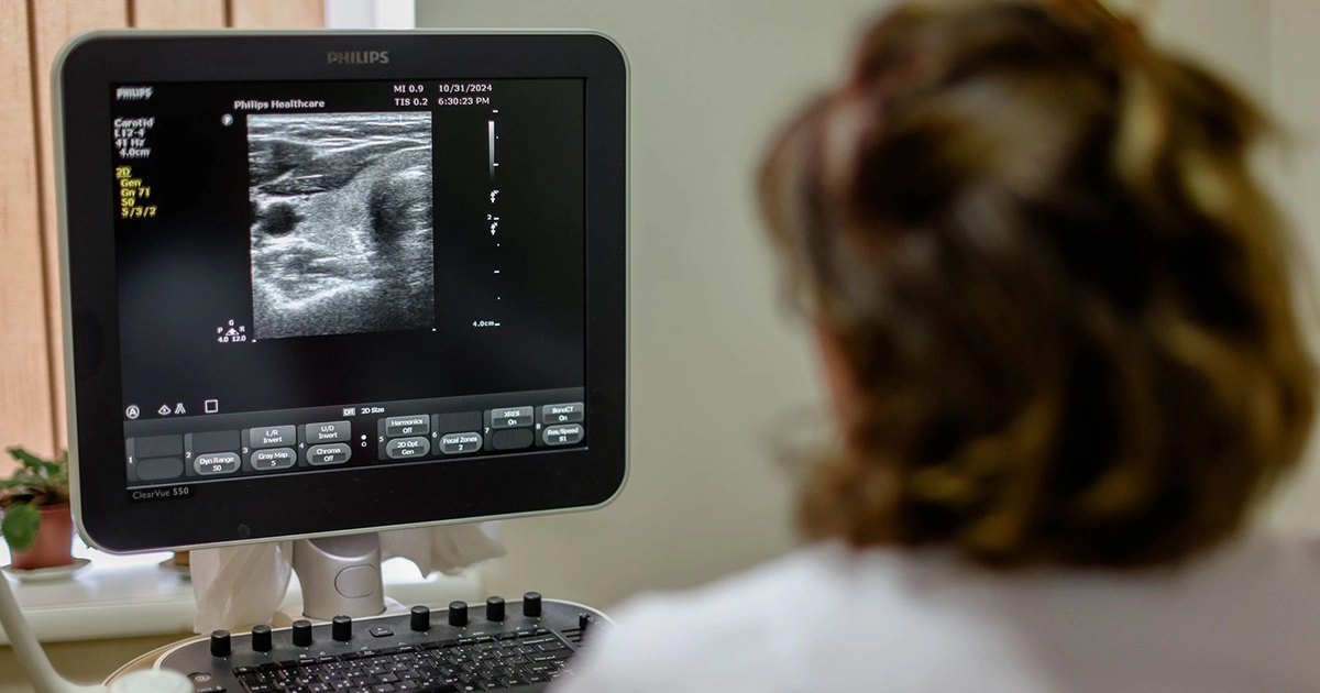
Ultrasonography machine functioning principle
Ultrasound is sound at a frequency that can’t be heard by a human ear. It passes through soft tissues and fluids, but is reflected as an echo from denser surfaces.
Higher frequency ensures a better image but is more easily absorbed by the skin and other tissues. Low-frequency sounds penetrate deeper into the tissues but the image of the object being examined is less sharp.
Ultrasound machine consists of 3 main parts: a control panel, display, and transducer probe that sends sound waves and receives reflected sounds (echo).
During the examination, the doctor moves a special sensor over the part of the body being examined, and the built-in computer analyzes the received signals, converts them into an image - a sonogram - and displays the image on the screen.
The shape and intensity of signals depend on tissue density. e.g. in the case of the fluid-fill cyst, most of the sound waves pass through this area, making weak reflection, therefore these areas appear as black areas. Ultrasound is well reflected by solid formations, creating a brighter and sharper image.
Ultrasound examination enables the study of the structure of organs and their condition at the stage of examination.
In addition, the method is used to monitor the biopsy process, when a small amount of fluid or tissue is removed for a laboratory study of the material properties.
Ultrasonography is also actively used for administering anesthetic and painkiller medications to topographically difficult areas.
Preparation for ultrasound examination
Preparation for ultrasonography examination take requires the consideration of the individuality and characteristics of the organ system being examined. This is necessary for good visualization of the organs so that the ultrasound examination can provide maximum information quickly and easily, to make a correct diagnosis. e.g.:
Therefore, when prescribing examination, the doctor explains the need to follow a special diet, if necessary, and other nuances.
The doctor should be necessarily informed about any medications you are taking
When coming for the ultrasound examination, it is reasonable to wear comfortable, loose clothes that don't hinder free movement and enable the patient to change positions simply, depending on examination nuances.
ICO is equipped with modern ultrasound machine (color Doppler), using which highly qualified specialists perform various types of examinations.
Dont miss Koi news


