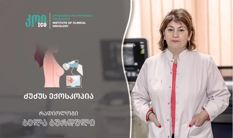
Breast echography (sonography) is a non-invasive ultrasound examination that enables a doctor to thoroughly examine breast tissue, milk ducts, nodules, and surrounding structures. This examination is especially important for both prevention and evaluation of specific symptoms, such as pain, swelling, or discharge.
When is a breast ultrasound prescribed?
A doctor prescribes an ultrasound in several cases, among them:
- A lump is found in the breast.
- There is pain and discomfort.
- Discharge from the breast.
- Change in the breast skin or shape.
- Post-treatment monitoring.
- Preventive purposes, especially in high-risk groups.
Breast ultrasound is often performed in younger women with denser breast tissue, when mammography is less informative. It is also used as a follow-up study to evaluate suspicious lesions detected by mammography.
How is the examination performed?
Examination is absolutely painless, requires no preparation, and lasts an average of 10-15 minutes.
What can a breast ultrasound detect?
Ultrasound can detect:
- Benign growths (cysts, fibroadenomas);
- Malignant tumors;
- Inflammatory processes;
- Enlarged ducts;
- Enlarged lymph nodes.
An ultrasound-guided biopsy can be performed if needed, as it is important for accurate diagnosis.
Advantages and safety
Breast ultrasound is completely safe for women of all ages. It can be done during pregnancy and breastfeeding. The exam can be repeated multiple times without any health risks.
When should it be performed?
It is recommended to have a breast ultrasound once a year for prevention in the 20–40-year age group, and to add a mammography after age 40. In high-risk groups (such as those with a hereditary background or genetic mutations), regular check-ups with a doctor and both examinations are necessary.
- Views:1424


