Radiographic mammography is a method of radiological examination of mammary glands, which enables the assessment of the breast image in a two-dimensional plane. This method is considered the “gold standard” for breast diagnosis, characterized by high (>92%) specificity.
Mammography with tomosynthesis is an in-depth study of the breast, during which we obtain both 2D and 3D images. During tomosynthesis, images of breast layers up to 1 mm thick are obtained, followed by the creation of a three-dimensional image from these layered images.
Tomosynthesis is especially informative in the case of dense breast glands (radiological density ACR3, ACR4) when pathology cannot be visualized on a standard mammogram due to a summative (caused by overlapping) image.
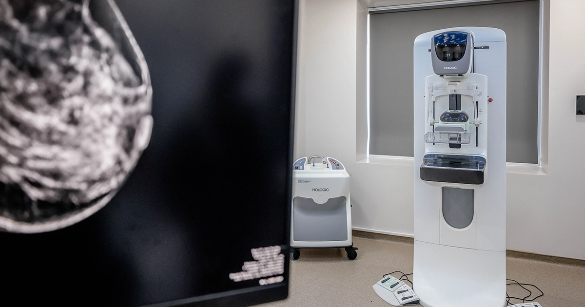
Standard mammogram shows a medial asymmetric density focus that may be assessed as Bi-RADS 3 (possibly malignant).
In tomosynthesis regimen, the neoplasm with unclear contours is exposed which is assessed as Bi-RADS 5 - highly suspicious malignant.
Multiple studies have shown that compared to a standard mammogram, tomosynthesis is 25-35% more effective in detecting breast cancer.
Benefits of tomosynthesis:

Dont miss Koi news


